See more and get top quality images with the MoleMax digital dermatoscope!
Now also the latest and most state-of-the-art MoleMax digital dermatoscopy device that includes unified mapping system of the moles is available in the Clinic of Dermatology and Surgery of Health Centre 4 (HC4) in Latvia. Besides it also provides possibility to examine pigmented newly formations onto the skin (moles, melanomas, basalioma etc.) in more details.
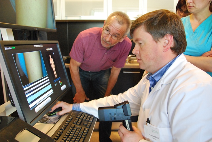
Representative of the DERMA MEDICAL SYSTEMS Josef A. Noesterer from Austria visited the Clinic of Dermatology and Surgery of Health Centre 4 (HC4); he provided an in-depth insight into the technological possibilities of the MoleMax and trained specialists of the HC4 to work with this new digital dermatoscopy device.

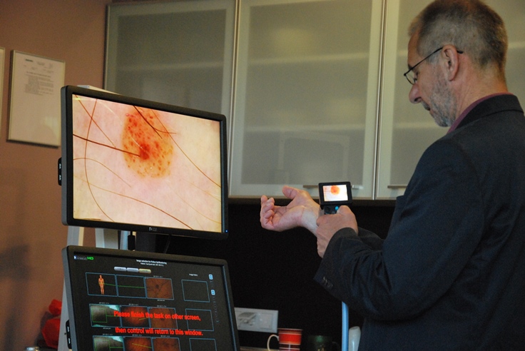
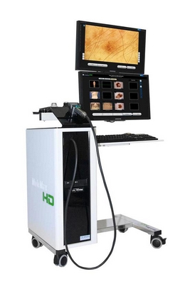
MoleMax device is a unique FULL HD TECHNOLOGY with two flat TFT monitors and additional video camera with an LCD screen for a preview. MoleMax technology provides possibility to obtain top quality high resolution image (it can be magnified up to 100 times), as well as this device features additional software for the evaluation of the image that allows not only precise analysis of the formations but also monitoring of their changes in a longer period of time. Device automatically compares all new formations registered in the system. Images that are pictured in a different period of time can be compared in real time obtaining valid comparison. Besides if the formation is removed, it is possible to add its histological image to the patient’s data base (case-record).
Device includes also an adjustable stand that fixates with 10 cm interval. This function allows to perform documentation of the whole surface of the body’s skin; this documentation is based on 33 separate segments (fast check-up comprises 10 segments). Due to the constant lightning it provides a high quality image from the top to toe like in the photo studio. Besides if you take pictures repeatedly you can obtain an image that is a precise analogue to the previous which was not possible before. In the mapping process of the formations it is very important to observe the moles in dynamics.

Josef A. Noesterer, sales manager of DERMA MEDICAL SYSTEMS, clinical trainer:
„I am delighted that now this technology is available also in Latvia. MoleMax device is a unique future technology that allows recognising suspicious formations onto the skin faster and observing them in dynamics in case of necessity, therefore, I hope that in the future this will reduce sickness rate and most importantly morbidity from the skin cancer. It is my second time in Latvia and I can surely say that the therapeutic care provided by the VC4 is of high level, especially, everything related to dermatology. Here different and latest technologies both in diagnostics and treatment are applied. As I have met doctors of this clinic for several times I have no doubts about the professional qualification as those specialists improve their knowledge in different congresses, workshops and trainings all around the world. They can surely be included in the group of, I am not afraid of this word, “world class” dermatoscopy experts”.
Raimonds Karls, dermatologist of the Clinic of Dermatology and Surgery:
I am satisfied about the entry of this new technology in our everyday work. This means that in this field we are progressing hand in hand with the experts of the world. And MoleMax technology is truly unique. First of all, patient who takes preventive steps to care for one’s health would benefit the most from the usage of this device in HC4. Device provides to the patient also visually understandable idea about his/her skin problems – nature of those skin formations. Second, this device facilitates work of the doctors as it is a unified and orderly system. Also MoleMax technology is ergonomically convenient to the doctor as you can connect to all software from the video camera but most important thing – it produces an image of exceptionally high quality and gives a chance to observe map of the patient’s moles to further observe those skin’s formation in dynamics. Therefore this examination and creation of the map of the moles is recommended to all people already having numerous formations in the skin and also children as the number of moles can increase from the age of 10 to approximately age of 40.”

